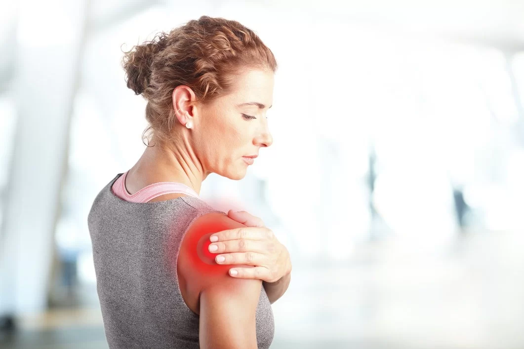
Shoulder pain is a problem that affects millions of people worldwide and can render the entire arm dysfunctional, making it difficult to perform daily activities.
The shoulders are prone to injuries, as they undertake the majority of the work in the rotational movements of the arms. Shoulder pain may occur at rest, during movement, worsen at night, or be constant. It may be accompanied by instability, stiffness, or muscle weakness.
Shoulder Pain / Risk Factors
Age and gender are risk factors for the onset of shoulder pain. It increases with age due to degenerative changes and occurs more frequently in women than in men.
Daily activities involving increased strain or repetitive motion—especially movements above shoulder level—during work (e.g., operating a jackhammer) or athletic activity (e.g., volleyball, basketball, javelin throw) are more commonly associated with the onset of pain.
Causes
Shoulder pain is caused by conditions and injuries of the shoulder joint, which may involve the bones, cartilage, or surrounding soft tissues (tendons, muscles, nerves). Less commonly, it may be referred pain from another part of the body (e.g., nerve compression in the cervical spine) radiating to the shoulder.
The most common causes include:
- Arthritis (osteoarthritis, rheumatoid arthritis)
- Tendinitis (inflammation of the tendon)
- Tendon rupture
- Dislocation (shoulder instability)
- Fractures
- Acromioclavicular joint disorders
- Inflammations
- Adhesive capsulitis, also known as “Frozen Shoulder” (mainly in women over 40)
- Calcific tendinitis
- Impingement syndromes
- Traumatic injuries
Diagnosis
Diagnosis is made through clinical examination and may require imaging tests, such as X-rays, MRI, or CT scans, to help ensure an accurate diagnosis.
Immediate management and treatment are essential, as the pain and associated mobility problems can worsen over time if left untreated.
Conservative Treatment
Initially, symptoms can be managed conservatively with:
- Resting the shoulder and, in some cases, using a brace to immobilize the arm and shoulder
- Applying cold packs (cryotherapy), and less commonly, warm packs to the affected area
- Taking painkillers and anti-inflammatory medications, as well as intra-articular or periarticular injections with hyaluronic acid or corticosteroids
At the same time, a targeted physiotherapy and muscle-strengthening program can be applied to improve range of motion and stabilize the shoulder. Additionally, in specific conditions, biological therapies using the patient’s own cells, such as PRP (Platelet-Rich Plasma) or stem cells, may be helpful.
Shoulder Arthroscopy
In cases where conservative treatment fails, arthroscopy is a minimally invasive and low-trauma method that allows the orthopedic surgeon to view the interior of the shoulder joint, identify, and treat any damage—usually without requiring hospitalization.
By inserting an arthroscope—a tiny camera—into the shoulder through a small incision (3–5 mm), the surgeon can:
- Carefully examine the inside of the joint and diagnose even very small or hard-to-reach injuries
- Repair the damage using specially designed micro-instruments and implantable inert materials, such as sutures and anchors
In most cases, arthroscopy takes about an hour, and the patient returns home the same day.
Different arthroscopic techniques are applied depending on the type and severity of the injury. When these do not help, minimally invasive shoulder arthroplasty provides a solution.
Minimally Invasive Shoulder Arthroplasty (M.I.S.)
Arthroplasty is a surgical procedure to replace damaged joint surfaces with highly durable, body-compatible implants. The main types of shoulder arthroplasty are:
- Total shoulder arthroplasty (replacement of the entire shoulder joint)
- Partial arthroplasty (replacement of part of the joint)
- Reverse total shoulder arthroplasty (replacing the entire joint with a special prosthesis that reverses the shoulder’s natural anatomy due to rotator cuff insufficiency)
Modern minimally invasive arthroplasty techniques involve much smaller incisions, use fast-track recovery protocols, and integrate digital technology to offer better and faster results than traditional methods.
Digital advancements allow the orthopedic surgeon to pre-plan the surgery in 3D, tailored to each patient’s unique shoulder anatomy, using a virtual environment. At the same time, the use of a navigation system or custom 3D-printed guides helps the surgeon perform the surgery with millimeter precision based on the digital plan.
The patient may be able to stand up just a few hours after the operation (once anesthesia wears off), begin moving the shoulder joint immediately, and return home within 1 day—depending on their clinical condition and medical history.
Physiotherapy
Whether the treatment is conservative or surgical, it is almost always followed by a rehabilitation program aimed at restoring the shoulder’s range of motion and strengthening the muscles. The duration of rehabilitation depends on the severity of the injuries and the patient’s functional demands.
ης αποκατάστασης εξαρτάται από τη βαρύτητα των βλαβών και τις λειτουργικές απαιτήσεις του ασθενούς.